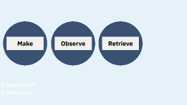
Competitive Landscape
Transcript: Competitive Landscape Microwells Competing Tech Large-scale companies Small-scale companies Make Competing Tech Similar Tech to Microwells In decreasing order of relevance: Nanowells and Micropatterend Surfaces Optofluidics Droplet Microfluidics FACS Micropatterned Surfaces / Nanowells Micropatterned Surfaces / Nanowells: similar to microwells, structured surfaces that trap cells or colonies Pros: can be combined with automated imaging and cell retrieval Companies: Bruker Optofluidics / Light-based Manipulation: Uses light to move cells into microchambers Pros: Very precise, allows functional assays + downstream recovery Cons: Costly, complex instrumentation Companies: Bruker Beacon, Lightcast Bio Optofluidics / Light-based Manipulation Droplet Microfluidics Droplet Microfluidics: cells are encapsulated, creating barcoded beads Pro: Good for genomics Limited live-cell imaging, and tracking function over time Companies: 10x Genomics, Mission Bio, Sphere Fluidics FACS FACS (Fluorescence-activated cell sorting):Classic technology, marker-based sorting of individual cells. Limitation: destructive, no longitudinal functional data, requires prior knowledge of markers. Large Companies Incorporating Microwells Large Competitors Thermo Fisher / Invitrogen – microwell plates (e.g., for spheroid/organoid culture). Corning – ultra-low attachment microwell plates for spheroid/organoid growth. Merck Millipore – makes microwell arrays for functional cell assays. 1CellBio – develops InDrop and other microwell-based single-cell analysis systems (though often combined with droplet methods). Nanocellect Biomedical – microwell + microfluidics hybrid for single-cell sorting. AIM Biotech – 3D cell culture chips with microwell arrays for tumor and immune-oncology studies. Cell Microsystems (CellRaft Arrays) – microwell-like “rafts” for single cell isolation, imaging, and retrieval. MicrowellDx (Scotland) – microwell-based molecular diagnostic assays. MIMETAS – organ-on-chip systems using microstructured wells for 3D cultures. InSphero – microwell plates for spheroid/organoid assays in oncology & toxicology. Cellix – microwell microfluidics for immune cell interaction assays. Startup Competitors Observe: Competitors offering live cell imaging Large-scale labs using live cell imaging Observe Single Cell imgaging in microfluidics Deepcell: high-throughput single-cell imaging + microfluidics; morphology analysis; uses AI Lightcast Bio (Envisia):single-cell functional assays (cell–cell interactions, killing); a cartridge/array microfluidic approach; uses ML analysis Sphere Fluidics (Cyto-Mine) NanoCellect (VERLO, WOLF) Mitrotisue and plate/chip live imaging InSphero MIMETAS AIM Biotech CytoSMART (now Axion BioSystems) Most Similar: Cell Microsystems (CellRaft / CellRaft AIR): microwell-style arrays for live imaging, clone tracking, then mechanical retrieval. AI: uses automated imaging + image analysis pipelines Phasefocus/Livecyte (acquired by Bruker): quantitative phase imaging (QPI) for single-cell tracking and long-term live assays (plate format). Live Cell Imaging Broad Institute — Imaging Platform & Assay Development — high-throughput live-cell imaging and image-based screening (Cell Painting, PERISCOPE, genome-wide imaging screens). Harvard Center for Biological Imaging (HCBI) — large imaging core offering live-cell imaging / high-content screening and training. MIT / Whitehead / Keck Microscopy Facility — broad microscopy cores with live-cell systems (spinning disk, confocal, intravital, timelapse). Stanford Cell Sciences Imaging Facility (CSIF) — campus core with live-cell confocal, high-content imaging and support. Large hospital cores — e.g., imaging cores at Boston Children’s Hospital, Dana-Farber/Harvard affiliates, Salk Institute, EMBL, Max Planck institutes also heavily use live-cell imaging for functional studies. Large-Scale Labs Retrieve Retrieval / isolation method AI/ML Live Cell Imaging? Yes Human Model + on-instrument AI for real-time extraction Deepcell (REM-I): Benchtop imaging + microfluidic/flow hybrid for morphology sorting Active sorting of viable cells into collection receptacles (6-way sorting). Yes automated image analysis pipelines and integrations with image tools Cell Microsystems (CellRaft AIR): Solid-phase array of releasable “cell rafts” imaged on-deck Mechanical release (needle) + magnetic retrieval → transfer to plates Yes “intelligent & adaptive” software for image/functional analysis Lightcast (Envisia): Droplet/cartridge benchtop platform that arrays + incubates single cells Cartridge dispensing/collection — allows for downstream follow-up. Yes On-instrument image/feature detection, supports downstream seletction Sphere Fluidics: Picodroplet encapsulation →incubation→on-chip imaging→dispense Automated isolation & dispense of hit droplets/cells into microtiter plates on-instrument feature extraction to guide gating & sorting Yes NanoCellect (VERLO): Microfluidic flow cytometry with integrated