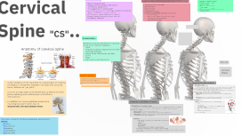
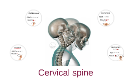
You're about to create your best presentation ever

Cervical Spine Powerpoint Template

Create your presentation by reusing a template from our community or transition your PowerPoint deck into a visually compelling Prezi presentation.

Cervical Spine

Transcript: suprascapular nerve Cervical Spine There are six cervical discs, because there is no disc between the upper two joints. The first disc is between the axis (C2) and C3. From this level downwards to the C7–T1 joint they link together and separate the vertebral bodies. Each is named after the vertebra that lies above: e.g. the C4 disc is the disc between the C4 and C5 vertebrae. Intervertebral discs lie between adjacent vertebrae in the spine. Each disc forms a cartilaginous joint to allow slight movement of the vertebrae, and acts as a ligament to hold the vertebrae together. Ligaments of the lower cervical spine The lesser occipital nerve zygapophysial joint or facet joint arises from the upper trunk (formed by the union of the fifth and sixth cervical nerves). It innervates the supraspinatus and infraspinatus muscles. Lower cervical spine A.Movements: flexion, extension, and lateral bending B.Bones: condyles of occipital bone and superior facets of atlas c.Ligaments and membranes : 1.Anterior atlanto-occipital membrane: membrane between the foramen magnum and the anterior arch of the atlas 2.Posterior atlanto-occipital membrane: membrane between the margin of the foramen magnum and posterior arch of the atlas 3.Lateral atlanto-occipital ligament: connects transverse processes of the atlas to jugular processes of the occipital bone The great auricular nerve forms the joint connecting the skull and spine. The atlas and axis are specialized to allow a greater range of motion than normal vertebrae. They are responsible for the nodding and rotation movements of the head.The atlanto-occipital joint allows the head to nod up and down on the vertebral column. The dens acts as a pivot that allows the atlas and attached head to rotate on the axis, side to side The anterior longitudinal ligament is closely attached to the vertebral bodies, but not to the discs. By contrast the posterior longitudinal ligament is firmly attached to the disc and is wider in the upper cervical spine than in the lower. Both ligaments are very strong stabilizers of the intervertebral joints. The lateral and posterior bony elements are connected by the ligamentum flavum, the intertransverse ligaments and interspinous ligaments and the supraspinous ligament or small occipital nerve is a cutaneous spinal nerve arising between the second and third cervical vertebrae, along with the greater occipital nerve. It innervates the scalp in the lateral area of the head posterior to the ear. The atlas and Axis second ligamentous complex connects the axis to the occiput: The cervical spine has seven vertebrae, which may be divided into two groups .The upper pair (C1 and C2, the atlas and axis) and the lower five (C3–C7) Anterior vertebral muscle Atlantoaxial Joint Atlanto-Occipital Joint Z-joint, or facet joint is a synovial joint between the superior articular process of one vertebra and the inferior articular process of the vertebra directly above it. The biomechanical function of each pair of facet joints is to guide and limit movement of the spinal motion segment. The lower cervical spine is composed of the third to the seventh vertebrae which are all very similar. The anteroinferior border of the vertebral body projects over the anterosuperior border of the lower vertebra. Intervertebral disc : Upper cervical spine muscles of posterior cervical B.Movement: rotation C.Bones: dens of axis articulates with the anterior arch of the atlas D.Ligaments 1.Anterior atlantoaxial ligament: from the body of the axis to the border of the anterior arch of the atlas 2.Posterior atlantoaxial ligament: from the lamina of the axis to the posterior arch of the atlas 3.Transverse ligament: spans across the arch of the atlas to hold dens against the anterior arch of the atlas originates from the cervical plexus, composed of branches of spinal nerves C2 and C3. It provides sensory innervation for the skin over parotid gland and mastoid process, and both surfaces of the outer ear Latral vertebral muscle

Cervical Spine

Transcript: 7 cervical vertebrae (C1-C7) that allow rotation of the head 6 intervertebral discs that act as shock absorbers and enable the neck to handle stress Facet joints allow small ranges of motion Anterior column: skeletal muscles Middle column: discs Posterior column: sensation Patients typically heal with proper treatment Pain should resolve Most common complication following fusion is decrease in neck motion Diagnosis of an Odontoid Fracture How does this injury change the normal function of the joint? Normal Anatomy The C2 part is injured. Aysha Ehsan and Caylee Vensel Which structure within the joint is injured? Treatment Prognosis Type II Odontoid Fractures occur when the cervical spine is hyperflexed (bent badly backward) or hyperextended (bent badly forward).Hyperflexion and hyperextension can be caused by trauma such as a fall or whiplash from a motor vehicle accident. Cervical Spine Type II Odontoid Fracture Computed tomography (CT): usage of special x-rays in order to view the organs, bones, and other tissues of a specific area in the body Examination: Neck pain gets worse with motion Dysphagia Myelopathy Break through C2 Numbness in back, arms, and legs Unable to rotate head When the Odontoid breaks,you cannot turn or twist your neck freely. How does it happen? The C2 is the axis;allows 50% of the heads rotation Nonsurgical care includes immobilization or restricted movement, or a cervical collar or halo vest Surgery is required if the fracture has resulted in neurological symptoms and the spine has become unstable Anterior screw fixation: enter from the front of the neck and place screws to hold the vertebrae together Posterior approach: C1 and C2 are fused together Sternocleidomastoid: Flexes cervical column to each side and elevates head by dorsally extending the upper cervical joints; expands thoracic cavity Sternohyoid: C1-C3; covers the front of the neck and lowers the neck and is involved in speech movement Interspinous ligament: connect spinal processes of adjacent vertebrae Transverse ligaments: connect lateral masses of the atlas and anchor the dens in place

Cervical Spine

Transcript: - Multi-level nerve root compression (tumour/space occupying lesion) - Myelopathy (spinal cord compression) - upper motor neuron lesions (MS, neurological conditions) - Cervical instability (potentially causing neurological compromise) - Cancer - Inflammatory arthrides - Congenital conditions - recent infection - deteriorating neurological status Cervical instability Caused by: - trauma - throat infection - congenital conditions - infalmmatory arhritides - surgery - head, neck, dental Recognition - lump in throat - unwilling to move head (bracing head) - metallic taste - you feel mm spasm/guarding ++ - symptoms change with movement Known Pathologies, Radiculopathy, Neuropathic pain Systems based approach Motor Control Dysfunction of the neck and associated areas Assessment Benign Paroxysmal Positional Vertigo - Nystagmus - Nausea/vomiting - Numbness/parathesia around mouth/nose area Range of motion Carotid Artery Dissection Combined movements Red flags Musculoskeletal neck pain Neurological Testing Mechano Sensitivity - ULNT VBI screen Posture 3 Ns - Dizziness - Diplopia - Double vision - Dysarthria/dysphasia – difficulty with speech - Dysphagia – difficult swallowing - Drop attacks – sudden collapse without loss of consciousness DDx 5 Ds Manual Assessment - PAIVMs - PPIVMs Vertebrobasilar Insufficiency Vascular Neurological Medical Conditions Musculoskeletal Conditions alignment and movement Articular nerve tissue neuromuscular sensorimotor Pain type nociceptive neuroplastic nociplastic Psychosocial Distress Anxiety Depression Fear Workplace Other

Cervical Spine

Transcript: What other pathologies are we concerned about in the cervical spine? Movement dysfunction Red Flags - Mrs Smith Motor control and sensory motor function Tone Warmth Trigger points Recommendations Atherosclerosis risk factors Hypertension Hypercholesteremia Smoker Palpation Tissue Mechanisms Manual therapy Hypertension - acutely raised BP can be a sign of vascular injury Hyercholesterolemia Hyperlipidemia Hyperhomocsyteinemia Diabetes Gentetic clotting disorders Infection Smoking Free radicals Direct vessel trauma Iatrogenic causes Palpatory techniques In Groups: Discuss what information we should gather to consider these pathologies The early presence of high levels of pain and disability, older age, cold hyperalgesia and moderate post-traumatic stress symptoms remain robust predictors of a poor outcome following whiplash injury at 2–3 years post-injury. (Sterling et al. 2006) Aetiology Covered in tissue and pain mechanisms But what are your thoughts about tissue mechanisms for Mrs Smith Patients with cervical pain/ WAD can present with deficits to their somatosensory/ or occulomotor system The following risk factors are associated with the potential for bony or ligamentous compromise of the upper cervical spine: History of trauma (e.g. whiplash, rugby neck injury) Throat infection Congenital collagenous compromise (e.g. syndromes: Down’s, Ehlers-Danlos, Grisel, Morquio) Inflammatory arthritides (e.g. rheumatoid arthritis, ankylosing spondylitis) Recent neck/head/dental surgery. Cervical instability describes a wide range of conditions from neck pain and deformation without any clear proof over little malformations too complete failure of intervertebral connection Psychosocial factors 1. What red flags does Mrs Smith present with? Daneel's planetary model of clinical reasoning in physiotherapy (2009) Vertebrobasilar insufficency 2. Are there any other factor which may not be red flags but need to be considered Kerry and Taylor (2006) explain "vertebral and carotid artery disease and dysfunction are intrinsically associated and two inter-related principles Underlying pathology (including atherosclerosis) may predispose a vessel to dissection Mechanical forces generated as a result of movement or biomechanics, can result in altered haemodynamics Pain Mechanisms Muscle length Think about quality as well as range - 60 years old - ? responsiveness to treatment - Dizziness Articular Myofascial Craniocervical flexion test Head Lift Global extension test Craniocervical extension test Typical cervical extension test Muscle strength/ co-ordination Upper cervical instability Flexion 40-45 Extension 45-70 Rotation 50-60 Lateral Flexion 20-45 Runs through the transverse forimina from C6-C1 Makes a sharp posterio-medial turn around the posterior mass of the atlas Is tethered to C1 and C2 Classic signs are 5Ds 3 Ns and ataxia 1. What red flags does Mrs Smith present with? 2. Are there any other factors which may not be red flags, but need to be considered? Exercise MOBS/ Manipulation PPVIMS PAVIMS Traction +/- neural treatment NAGS/ SNAGS Active or self treatment PIN principle ROM exercise Massage Trigger point release MET Myofascial release PAIVMS Useful for identification of painful segments PPIVMS Useful for identification of movement restriction Think about potential adjustment which may aid differentiation The ICA provides the most significant proportion of blood to the brain Pathological changes to the ICA are very common Blood flow in the ICA is known to be affected by cervical movmeents What is the effect you are looking to achieve? Pain relief/ change in range Grading to match goal Be Suspicious Pain may be the first presenting sign Consider ICA as well as VBI Think about cardiovascular risk factors and testing blood pressure If confident utilise cranial nerve testing When frank acute injury is suspected A&E. If more chronic presentation recommend urgent referral to BICS with indication for US doppler Scalene Anterior Middle Posterior Upper fibres of traps Levator Scapula Pec major and minor Neurogenic - Not going to be discussed due to neuro in-service Cervical Artery Dysfunction Range of motion Cervical Spine Junior In-service March 2014 Stretches Strengthening Proprioceptive Self SNAGS Thank you for your time... Any last questions??? Types of dysfunctions include: Somatosensory dysfunction Disturbed head neck awareness Disturbed neck control Disturbed postural stability Occulomotor dysfunction Smooth pursuit Gaze stability Saccadic movement Eye head co-ordination Subjective and objective signs that indicate the presence of serious pathology Internal Carotid Artery Treatment for articular dysfunction Quebec Task force 2008 Post injury psychological distress Passive types of coping Compensation Look at active and passive

Cervical Spine

Transcript: Injuries from the atlantoaxial joint can vary from being able to fully recover to fatality. Sprains and strains... Rectus Capitis Lateralis : Originates on the upper surface of the transverse process of the atlas and inserts under the surface of the jugular process of the occipital bone. Problems Materials Obliquus Capitis Superior: Originates on the lateral mass of the atlas and inserts on the lateral half of the inferior nuchal line. Intertransversarii Anteriores Posteriores: Originates on the transverse process and inserts on the transverse process above. Cervical Herniated Discs... It is responsible for the majority of the heads flexion and extension mouvments. The Atlantoaxial joint is a slightly movable or cartiliginous joint. The Atlantoaxial Joint Some difficulties we had with building our joint: -Finding out where everything attaches - Finding the right materials to represent the joint and that wouldn't fall apart Muscles and Tendons In conclusion... When the the upper body is hit or moved suddenly it can cause a neck sprain or strain to occur. This can also cause a hyper extension when your head goes backwards, if this occurs quickly or with force any of these movements can cause a sprain or strain. The bones involved in this joint are the axis (C2) and atlas (C1). Rectus Capitis Anterior : Originates on the atlas and inserts on the basilar part of the occipital bone. Obliquus Capitis Inferior: Originates on the spinous process of the atlas ans inserts on the lateral mass of the atlas. Sports Injuries This occurs when the discs that line each vertebra become tears this is often because of overuse. Neck pain is usually the first symptom but you can also experience pain throughout your arm. Fractured Neck The atlantoaxial joint is a very important joint that is used for everything that you do and should be taken care and protected while participating in sports. paint plasticine a pipe glue elastics fabric elastics ball pin paper The atlantoaxial is a pivot joint. The movements that it is able to do is rotations. By Miia and Sarah Sports Injuries To construct our model of the cervical spine we used: The atlantoaxial joint is apart of the cervical spine, and more commonly know as the neck. The cervical spine is more mobile than the thoracic and lumbar regions. Muscles and Tendons Atlantoaxial Joint Caused by a severe blow to the head or neck or a sudden twist in the neck. Common Sports this can happen in are football, hockey, rugby, wrestling, diving, and skiing. Neck injuries are most likely serious as they often relate to the head and nervous system. The tendons and muscles involved in the atlantoaxial joint are: Atlantoaxial Joint Some of these injuries include:

Cervical Spine

Transcript: Suggested Protocol - GE 1.5 Tesla 3 plane localizer SAG T2FSE SAG T1 FSE Axial T2 Axial T1 STIR Precontrast: Postcontrast: References Grey, M., Ailinani, J. (2012). CT and MRI Pathology: A Pocket Atlas. (2nd ed., p. 104). The McGraw-Hill Companies. Conclusion Enhancement was seen within lesion in pre-contrast and post- contrast images. Also small area of increased signal has developed in the cord to the left of midline at the C5 level. These changes are consistent with MS plaques. Multiple sclerosis is demyelinating disease affecting the spinal cord and the brain tissues. MS has periods of exacerbation and remission. In this case, the size of the lesion has increased from 1cm to 2 cm when compared to the previous MRI study. This means that patient underwent exacerbation period. There is no specific treatment for MS. Corticosteroids and other drugs, however, are used to treat the symptoms. Also therapy may help to postpone or prevent specific disabilities. Further MRI studies will be helpful in monitoring the disease progression. Findings and Discussion Patient history The MRI technologist explained to the patient the process of the MRI exam. MRI technologist went over screening form with the patient to make sure that the patient does not have any unsafe MR metals in her body. Then the patient was instructed to change her clothes and remove all jewelry. The patient was positioned supine in CTL coil. MRI tech provided cushion under her knees for comfort and offered a warm blanket. In addition she was provided with ear plugs and call button. The patient was landmarked at the middle neck area. She was scanned using 1.5 T GE magnet machine. Before leaving the room, technologist double checked that the patient is able to communicate. The door was secured, shut and study began. A 32- year- old female presented to the hospital with left sided weakness. A magnetic resonance imaging (MRI) was obtained. The study was ordered with contrast. Lab work indicated that the GFR of this patient was > 65. The patient has no history of injury and surgery. This patient had MRI study completed three years ago and was diagnosed with Multiple sclerosis of spinal cord. At that time the size of the lesion was 1 cm. Cervical Spine Multiple Sclerosis (Spinal Cord) Patient Preparation and Scan Set-up SAG T1 Axial T1

Cervical Spine

Transcript: The father, Atlas (aka C1) sits at the base of the skull. He has the largest opening out of all of us because this is where he and the brain and/or skull meet up. He ALWAYS says "Yes" Our bodies are all made out of the same parts. We have a vertebral body, pedicle bones, lamina bones, spinous processes, transverse processes, and transverse foramen. We also have a lot of responsibility for the rotation, flexion, and extention of the head and neck! Mom and Dad are in charge of the head movement while my younger 3 brothers (C5, C6, C7) are in charge of flexion and extention! Conclusion....... DONE? Some problems our family can have: Other conditions: Fractures and spinal cord injuries that can debilitate, paralyze or cause death. Not good and needs to be taken seriously! We are made up of 7 family members Whiplash: sudden jerking motion to the head (a direct blow) causing head to violently jerk back and forth. I am the C-Spine Family Disc Herniation: pain, spasms, weakness, numbness, and some possible paresthesia. Common in my brothers C6 and C7. The mother, Axis (C2) has a pointy know called the dens that sits up into the Atlas. Mom always tells us "No" On the sides of us we have transverse processes with a hole called transverse foramen. This is where arteries supply blood throughout our whole neighborhood to keep the it all alive and well. Cervical Degeneration: cervical spurs or cerivical osteoarthritis. Cervical Osteoarthritis is the breakdown of cartilage inbetween the facet joints in our "C-spine" family. Pain presents early mornings, late nights, and calms with rest. Could cause pain in shoulders or shoulder blades. most common, stingers or burners (compression of the nerve root usually between my brothers (C5-C7). This can cause numbness, tingling, maybe some weakness to the extremities. This could take minutes to hours to resolve. Common reoccuring event. We all have an important job together as a family, this includes: Housing and protecting the spinal cord. supporting our neighbor, the head. Also facilitating blood flow to the brain.

Now you can make any subject more engaging and memorable