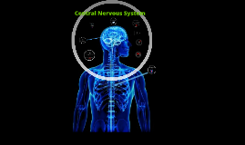
Central Nervous System
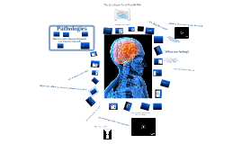
Transcript: What is it? *composed of brain and spinal cord and is made up of cells called neurons Make-Up: Frontal Lobe- responsible for thought, language, emotions, and voluntary movements Parietal Lobe- responsible for perception and interpretation of touch Occipotial Lobe- where visual images are processed Temporal Lobe- recognized and interprets sounds and helps form new memories Colliculi- relays visual and auditory sensations throughout the cerebrum Functions *allows communication to the body and responds to stimuli *transmits nerve impulses from all parts of the body *controls virtually everthing (I.E. sight, smell, hearing, taste, feeling, emotion) *carries electrical signals and transmits them to other nervous cells, muscular cells, and glandular cells Breakdowns *Nervous system problems may occur slowly and cause a gradual loss of function (degenerative), or they may occur suddenly and cause life-threatening problems (acute) Some causes of nervous system problems: *vascular disorders *trauma, especially injuries to the head and spinal cord *congenital problems (are present at birth) *anxiety disorders, depression, or psychosis *exposure to toxins, such as carbon monoxide, arsenic, or lead -Problems that cause a gradual loss of function (degenerative): *Parkinson's disease *Multiple sclerosis (MS) *Alzheimer's disease *Amyotrophic lateral sclerosis (ALS) *Huntington's disease *Peripheral neuropathies -Infections: *brain: encephalitis or abcesses *membrane surrounding the brain and spinal cord: meningitis Quiz Time True or False: The central nervous system is mainly made up of the brain and heart. False; It's mainly made up of the brain and spinal cord. True or False: The central nervous system only controls your sense of smell and taste. False; it controls sight, smell, hearing, taste, feeling, emotion, and much more. Which of the following will help maintain your central nervous system's health? a. eat a lot of chocolate b. eat healthy fats c. write as neatly as possible for 15 minutes a day d. a & b e. b & c e. b & c Nervous system problems are... a. degenerative b. acute c. a or b d. none of the above c. a or b True or False: The central nervous system carries electrical signals and transmits them to other nervous, muscular, and grandular cells. True Works Cited Batigne, Stephane, Josee Bourbonniere, and Nathalie Fredette. Major Systems of the Body. MIlwaukee, WI: World Alminac Library, 2002. Print. Macaulay, David. The Way We Work. New York City, NY: Hofton MIfflin Company, 2008. Print. MD, WEB. “ Nervous System Problems.” WedMD. WedMD, Inc., 2012. Web. 6 Feb. 2012. <http://www.webmd.com/brain/tc/nervous-system-problems-topic-overview>. *make sure you intake adequate amounts of healthy fats, vitamin D, and vitamin B12 *spend a minimum of 15 minutes per day writing on paper as neatly as you can (writing requires the use of all major components of your conscious motor and sensory pathways) Maintain It's Health! Central Nervous System