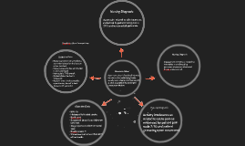
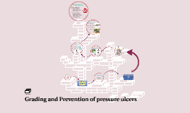
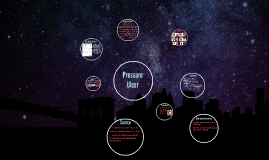
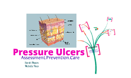
You're about to create your best presentation ever

Pressure Ulcer Powerpoint Template

Create your presentation by reusing a template from our community or transition your PowerPoint deck into a visually compelling Prezi presentation.

Pressure Ulcer

Transcript: Unstageable Skin damage in which there is full thickness skin and tissue loss whereby the extent of tissue damage is unconfirmed because it is obscured by slough or eschar. Identify At Risk Patients Prevention Stage 1 Observable pressure related changes of intact skin You will find: - Skin temperature changes (warm or cool) - Tissue consistency (firm or boggy) - Sensation (Pain/Itching) - Appearance of lesion ->> Defined area of persistent redness in lightly pigmented skin or persistent Red/blue or purple in darker skinned -> Identify at risk patients -> Pressure redistribution ->> Supportive surfaces ->> Position and inclination ->> Repositioning -> Supportive interventions ->> Improve mobility ->> Improve skin perfusion ->>Provide adequate skin care ->> Minimize moisture ->> Correct Malnutrition Grading- According to NPUAP Bed or chair bound Poorly fitting casts Malnourished Decreased blood flow to the skin Reduced sensation to the skin People at risk Stage 3 Full thickness skin loss involving damage or necrosis to subcutaneous tissue that may extend down to but not through underlying fascia. Presents clinically as: -> A deep crater with or without erosion of adjacent tissue Supportive Interventions Pressure Redistribution Improve Mobility Improve skin perfusion Provide adequate skin care Correct malnutrition Stage 4 Full thickness skin loss with extensive destruction, tissue necrosis or damage to muscle, bone or supporting structures e.g tendons or joint capsule. Erosion and tunneling/sinus tracts often occur Grading and Prevention of pressure ulcers Pressure Ulcers Stage 2 Partial thickness skin loss involving the epidermis and or dermis - Appearance ->> Superficial lesion that presents as either and abrasion or blister Supportive surfaces Position and inclination Regular repositioning- every 2-6 Hours A pressure ulcer is a localised injury to the skin and or underlying tissue occurring over the bony prominences as a result of pressure or pressure in combination with shear and or friction! The most cost effective way of managing pressure ulcers is PREVENTION !! add logo here

Pressure Ulcer

Transcript: suspected deep tissue injury. Also known as bedsores or decubitus ulcers Symptoms/Tests Stages Source Stage 2 Pressure Ulcer 10% to 18% in acute care, 2.3% to 28% in long-term care, and 0%-29% in home care Treatment Stage 4: The pressure ulcer has become so deep that there is demage to the muscle and bone, and sometimes to tendons and joints. www.nlm.nih.gov/medlineplus/pressuresores.html Stage 3 Deep Tissue Injury: the skin is purple or maroon-red, appears as a blood-filled blister. as of 2010, pressure ulcers resulted in about 43,000 deaths. Stage 1: A reddened area on the skin that,when pressed, does not turn white.- sign of ulcer forming. The effected skin looks red and my feel warm to touch. it may also burn, hurt or itch. Red skin that gets worse over time The area forms a blisterc then an open sore Tests- Blood Test, Tissue Cultures (diagnose a bacterial or fungal infection, also for cancerous tissue A pressure ulcer is an area of skin that is damaged from staying in a position for to long. Pressure ulcers form on your ankles, back, elbows, heels, hips, butt, shoulders, and at the back of your head. Prevention Causes Stage 2: The skin blisters or forms an open sore. the area around the sore may be red and irritated. Stage 3: the skin now develops an open, sunken hole or crater. The tissue below the skin is damaged. Change position at least every 2 hr to relieve pressure. Eat well-balanced meals that contain enough calories to keep you healthy. Drink plenty of water every day. Exercise daily Keep the skin clean and dry www.mayoclinic.com/health/bedsores/ds00570/dsection=symptoms umm.edu/health/medical/ency/articles/pressure-ulcer Relieve the pressure on that area. Treat the sore avoid further injury or friction to the area. Eat healthy foods. Clean the ulcer the way your doctor or nurse told you to. Rinsed with a salt-water- removes looes, dead tissue. Stage 1 Unstageable pressure ulcers: The damage can't be estimated. www.logicalimages.com/publichealthresources/pressureulcer.htm Stage 4 The pressure on the skin reduces blood flow to the area. Without enough blood, the skin can die and an ulcer may form. Also staying in bed for a long time can cause ulcers. People with fragile skin and paralyzed people have a higher chance of getting ulcers. Additional Information unstageable pressure ulcers

Pressure Ulcer

Transcript: Pressure ulcers occur most commonly over a bony prominences Pressure ulcers may develop on ANY part of the body under a splint or cast external fixations devices tubes pulse oxymetery meters Full thickness tissue loss with exposed bone, tendon or muscle, tissue necrosis, or damage to muscle, bone, or supporting structures; tendons and joints Objectives GEL PADS FOR EVERYONE! Pressure Ulcer Care Lewis, S. L., Dirksen, S. R., Heitkemper, M. M., & Bucher, L., & Camera, I. M. (2011). Medical-surgical nursing: Assessment and management of clinical problems (8th ed.). Maryland Heights, MO: Elsevier. Mayo Clinic, (2001). Bedsores (Pressure Sores) Prevention. Mayo clinic. Retrieved from: http://www.mayoclinic.com/health/bedsores/DS00570/DSECTION=prevention The NPUAP, (2013). Pressure Ucler category/staging illustrations. The National Presure ucler Advisory Panel. Retrieved from: http://www.npuap.org/resources/educational-and-clinical-resources/pressure-ulcer-categorystaging-illustrations/ POSITION, POSITION, POSITION!!! Thank you! How often should you turn a patient to prevent pressure Ulcers? What stage is an ulcer when you can see fatty tissuse but no bone or muscle? Name two risk factors for developing a pressure ulcer? Partial thickness loss of epidermis and dermis. The ulcer is superficial and appears as an abrasion, blister, or a shallow crater. A shiny or dry shallow ulcer without slough or bruising Patients older than 65 years old Diabetes Mellitus Edema Vasoconstrictive diseases Stage 4 Stages of Pressure Ulcer Inspect integrity of skin especially under and around any protective devices used Use proper turning and transfer techniques and devices to avoid sliding friction and shear Document any skin discrepancies and report, take a picture! Contact wound care specialist to inspect and treat the ucler Regularly inspect and clean skin in contact with probes, drains ,cannula and tubing. Provide patient with proper nutrition Stage 2 incontinence Skin discomfort( pain, Itching, loss of sensation) Skin blanching, abscess or absence of skin layers Care Tissue tolerance- The integrity of the skin Shear – occurs when the patient slides downwards with gravity while the skin remains in the original position, stretching and tearing the blood vessels causing tissue ischemia. Friction- occurs when two surfaces moves across each other removing superficial layers of the skin usually over a bony prominence. Gel pad arm on arm boards to relieve pressure on elbows Gel pad on the heels of patients in suspension slings Gel pad head ring to protect occipital area Pillows to protect boney promiences Prevention of Pressure Ulcers Full thickness tissue loss. Subcutaneous fat may be visible but bone, tendon or muscle are not exposed. Risk Factors 3 Keep patient dry with no creases under patients skin Change patient position every 2 hours Apply the appropriate topical agent or dressing in relation to the ulcer characteristics as ordered. Reassess skin to check for any change in skin integrity Pressure Points Skin hygiene Skin moisture Skin temperature Skin integrity Skin scarring REFERENCEs If possible re-position the patient every 2 hours to relieve pressure from the affected areas Avoid high semi-fowlers position for extended periods as it increases the shear and pressure forces in the sacral area. Position high risk patients on specialty beds if possible Intact skin with non-blanchable redness/erythema of intact skin of a localized area usually over a bony prominence. Discoloration of the skin, warmth, edema, hardness may also be used as indicators, particularly on individual with darker skin. Stage 3 jk QUIZ 1 OTHER PRECAUTIONS Sarah Mason Michelle Rees Avoid pooling of fluids and/or secretions under patients keep skin dry at all times Take special care of blood pressure cuff areas and rotate site if necessary Sheets, patient gown and linen must have no creases or folds beneath patient Stage 1 A pressure ulcer is an area of localized damage to the skin and underlying tissue that is caused by pressure, shear, friction and or a combination of these. Identification of patients risk of developing pressure ulcers How to Identify the stages of Ulcers How to prevent Ulcer formation Pressure Ulcers Assessment.Prevention.Care Factors in the formation of pressure ulcers Pressure Ulcer 2

Pressure ulcer

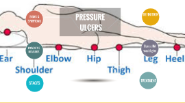
Transcript: stage4 stage 3 NHS reported that just under 25000 reproted to develop pressure ulcer from 2014 -205 20% of people in acute care & 30% of people in community at risk. pressure ulcer treatment is a costly process . The UK spends around 1.4 to 2.1 billion yearly. (Bennett, at al,. 2004). objective prevention pressure ulcer mostly occur as a result of inadequate care, following the principle of prevention help protecting people at risk from developing pressure ulcer Nonblanchable erythema of intact skin heralding lesion of skin ulceration. In individuals with darker skin, discoloration of the skin, warmth, oedema, induration or hardness may be indicators. kareema alabri References Thank you mobility problem poor nutrition underlying health condition being over 70 years old incontinence : urine/ bowel serious mental health condition Full thickness skin loss involving damage to or necrosis of subcutaneous tissue that may extend down to, but not through underlying fascia. The ulcer presents clinically as deep crater with or without undermining of adjacent tissue (Ellis, 2016) How it occurs The extra pressure disrupts the blood flow through the skin. Without the blood supply, the effected skin areas became starving of oxygen and nutritions.it began to breakdown leads to formation of ulcer which can be infected by bacteria. stage 2 content stages 1 areas of damaged skin and tissue that developed from sustained pressure usually laying in bed or sitting in wheelchairs. Range in severity from patches of discoloration skin to open wounds that expose the underlying muscle or bone . (Morison, 2001) (Ellis, 2016) Partial thickness skin loss involving epidermis, dermis, or both. The ulcer is superficial and presents clinically as an abrasion, blister, or shallow canter. (Ellis, 2016) sites (Keller, et al., 2002) At the end of this session the student will be able to define pressure ulcer. states the causes and how it occur. identify the sites and stages. list the measures to prevent it. (Ellis, 2016) Toward reducing the pressure ulcer incident throw implementing the prevention measurement. At risk Full thickness skin loss with extensive destruction, tissue necrosis, or damage to muscle, bone, or supporting structures (e.g, tendon, joint capsule). Undermining and sinus tracts also may be associated with Stage IV pressure ulcers. (NHS Choices information) Pressure ulcer Conclusion What is pressure ulcer How it happen At risk Sites and stages Braden scale Prevention Bennett, G., Dealey, C. and Posnett, J., 2004. The cost of pressure ulcers in the UK. Age and ageing, 33(3), pp.230-235. Bergstrom, N., 1994. Treatment of pressure ulcers (No. 95). DIANE Publishing. Cannon, B, & Cannon, J 2004, 'Management of pressure ulcers', American Journal Of Health-System Pharmacy, 61, 18, p. 1895, Academic Search Premier, EBSCOhost, viewed 24 November 2016. Lyder, C.H., 2003. Pressure ulcer prevention and management. Jama, 289(2), pp.223-226. Langer, G., Knerr, A., Kuss, O., Behrens, J. and Schlömer, G.J., 2003. Nutritional interventions for preventing and treating pressure ulcers. The Cochrane Library. Bergstrom, N., Braden, B.J., Laguzza, A. and HOLMAN, V., 1987. The Braden scale for predicting pressure sore risk. Nursing research, 36(4), pp.205-210. House, K.W. and Johnson, T.M., 2014. Prevention of Pressure Ulcers. In Pressure Ulcers in the Aging Population (pp. 27-45). Humana Press. Morison, M. J. (2001) The prevention and treatment of pressure ulcers. London: Mosby. Ellis, M 2016, 'Understanding the latest guidance on pressure ulcer prevention', Journal Of Community Nursing, 30, 4, pp. 29-36, CINAHL Complete, EBSCOhost, viewed 25 November 2016. https://i.ytimg.com/vi/9AvCUJgT2zI/hqdefault.jpg (accessed on 7/12/2016). http://www.nhs.uk/Conditions/Pressure-ulcers/Pages/Causes.aspx, (accessed on 7/12/2016). Keller, P.B., Wille, J., van Ramshorst, B. and van der Werken, C., 2002. Pressure ulcers in intensive care patients: a review of risks and prevention. Intensive care medicine, 28(10), pp.1379-1388. What is pressure ulcer? Aim

Now you can make any subject more engaging and memorable