Foot
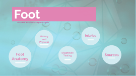
Transcript: Chana Rich, Neha Raukar, and Jhanavi Kapadia Foot Foot Anatomy The foot is made up of 28 bones and 57 articulations. It can be divided into three regions: the hindfoot, midfoot, and forefoot. Hindfoot- talus and calcaneus Subtalar joint-comprised of three articulations between the talus and calcaneus Midtarsal (Chopart's) joint connects the hindfoot to the midfoot Midfoot- navicular, cuboid, and cuneiforms (medial, intermediate, lateral) Tarsometatarsal (Lisfranc's) joint connects the midfoot and forefoot Forefoot- metatarsals, phalanges, and sesamoids. The plantar fascia maintains the arches Vascular anatomy- The arterial supply to the foot is from the anterior and posterior tibial arteries and peroneal artery, a proximal branch of the posterior tibial artery. Nerves- Motor and sensory innervation comes from branches of the deep and superficial peroneal, posterior tibial, saphenous, and sural nerves Foot Anatomy Bones https://upload.wikimedia.org/wikipedia/commons/5/52/812_Bones_of_the_Foot.jpg Anatomy Review: Skip to 0:45 History and Physical History and Physical History If the patient remembers, ask about the mechanism of injury. It is important to ask about the timing and duration of symptoms. Additional helpful information includes the location of pain, the description of its quality, and precipitants. Underlying medical conditions and medications which would impede on wound or fracture healing are important. Don’t forget to ask about previous foot problems. History Physical Exam Assess the foot in its position of rest (slight plantar flexion and inversion) Evaluate for swelling, deformity, ecchymosis, open wounds, color, and temperature Palpate for any areas of tenderness Palpate a dorsalis pedis artery pulse Check sensation in all nerve distributions (see neuroanatomy section) Evaluation active and passive range of motion Observe the patient while standing-do you notice a high arch or a flat foot? Any bunions? Observe gait Physical Exam Quick foot exam review: https://www.youtube.com/watch?v=Bbt2I2ZE9uA Diagnostic Testing Diagnostic Testing Ottowa Foot Rule Ottowa Foot Rule Ottowa foot rule state that a foot radiographic series is required if there is pain in the midfoot region with any of the following findings: Bone tenderness at the navicular bone Bone tenderness at the base of the fifth metatarsal Inability to bear weight for at least four steps immediately after the injury and at the time of evaluation Imaging X-rays: Standard three-view radiographs of the foot consist of anteroposterior, lateral, and oblique projections. Foot radiographs are difficult to interpret. Additional views, calcaneus views or weight-bearing views may be indicated. Accessory ossification centers are seen in 30% of the population Unlike fractures, they have smooth corticated surfaces CT scan: helpful when a fracture is suspected, but x-rays are unrevealing. CT is particularly helpful when imaging the midfoot, calcaneus, subtalar joint and tarsometatarsal (Lisfranc) joint. Imaging Normal X-Rays https://commons.wikimedia.org/wiki/File:X-ray_foot_1.jpg https://www.youtube.com/watch?v=XYkVUrsYKes CT Scan Fractures Injuries Hindfoot Fractures Hindfoot Fractures These fractures can be missed as the presentation can be similar to an ankle sprain. Talur Fractures Talar Fractures: The talus has no muscular or tendinous attachments, but it does have 7 articulations. The blood supply to the talus can be disrupted by fractures or dislocations, putting the talus at risk of avascular necrosis. Talar Neck Fractures: most common fracture of talus (50%). This is the only part of the talus that is extraarticular. This is caused by a high-energy mechanism, usually axial load or dorsiflexion. All cases require emergent closed reduction in ED. Nondisplaced fractures can be treated with immobilization and non-weight bearing for 6 weeks. All displaced fractures are operative. The Hawkins Classification is used to describe talar neck fractures with type I being a non-displaced fracture and type IV being a subtalar, tibiotalar, and talonavicular dislocation. Hawkins IV fractures have up to a 100% chance of avascular necrosis of the talus. Talar Body Fracture: Articulates with the tibia, fibula and calcaneous. Lateral Process Fractures: Often seen in snowboarders Talar Head Fracture: least common. The head articulates with the navicular and calcaneus. Talar head fractures almost always involve the talonavicular joint, and associated dislocation is common. Subtalar Dislocation: Simultaneous disruption of the talocalcaneal and talonavicular joints. Subtalar dislocation is described by the direction of the foot in relation to the talus. Medial dislocations are the most common. Patients typically have an obvious deformity on exam. These should be reduced right away by flexing the knee and applying longitudinal traction to the foot with initial accentuation, followed by reversal of the deformity. Following reduction, immobilize with a short leg cast for